DIFFERENCES AMONG NEURONS
THE NEURON
WHAT IS A NEURON?
STRUCTURE OF THE NEURON
DIFFERENCES AMONG NEURONS
THE SYNAPSE
NERVE IMPULSES
PROCESSES OF EXCITATION AND INHIBITION
SUMMARY
BRAD JORGENSEN
B.S.Jorgensen@bath.ac.uk
EXT: 6851
WHAT IS A NEURON?
Nerve cells or Neurons are cells that exist in the nervous system - they are the basic building blocks of the nervous system.
The nervous system is like a large communication network, and consists of two major parts:
PERIPHERAL NERVOUS SYSTEM (PNS) - Acts like a transmitter (i.e., input-output). It contains neurons that either (1) gather information from the environment, or (2) deliver information to muscles and glands.
CENTRAL NERVOUS SYSTEM (CNS) - Consists of neurons found in the brain and spinal cord. The CNS processes incoming information and issues instructions that underlie the way we respond.
There are in excess of 100 billion neurons in the
nervous system, with most being located in the brain. Each neuron is connected
with thousands of others in a complex network of neural connections.
NEURONS VERSUS NON-NEURAL CELLS IN THE NERVOUS SYSTEM
Glial cells are non-neural cells that are not involved in the process of transmitting information in the nervous system. They perform a number of important functions:
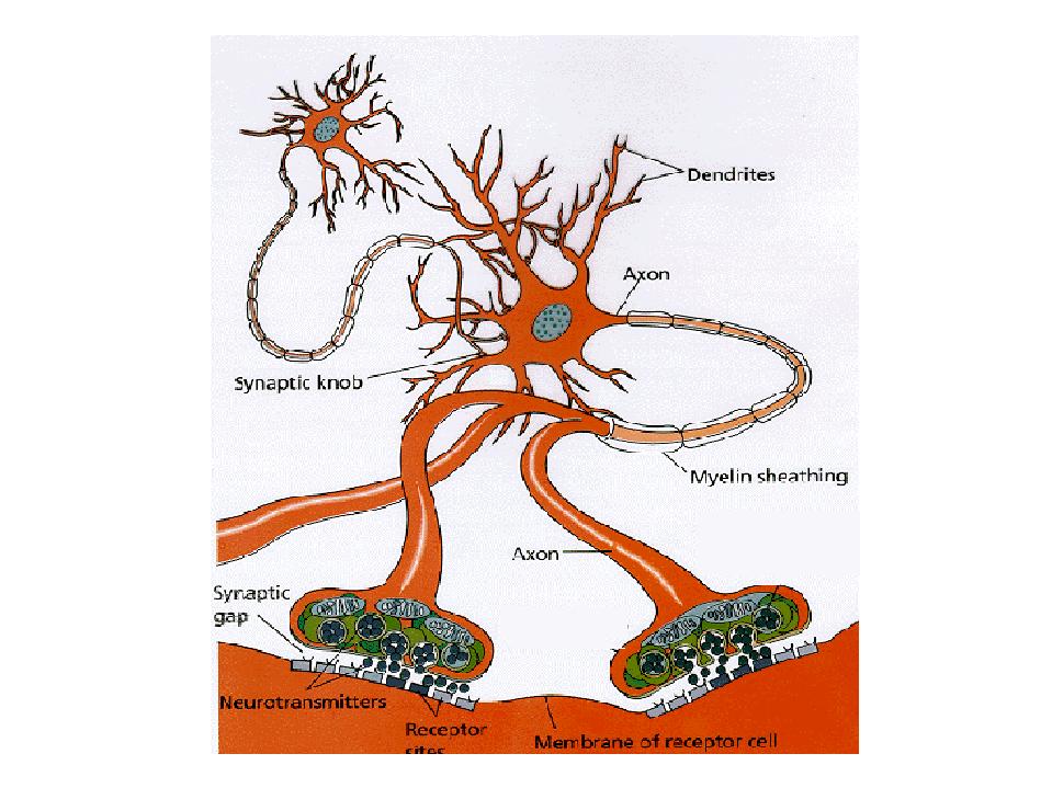
STRUCTURE OF THE NEURON
As the basic operating unit in the nervous system, the neuron functions to transmit information from one part of the body to another. This information takes the form of an electrochemical signal called a neural impulse.
SOMA (or CELL BODY) - place where the neuron processes nutrients into energy.
NUCLEUS - a round or oval body that contains the genetic information necessary for the control of cell structure and function.
DENDRITES - multibranching extensions of the neuron that receive information from other cells.
AXON - long, single extension of the soma that forms the communication line of the neuron.
AXONAL ENDINGS - the branches located at the end of the axon that make connections with muscles or other neurons.
MYELIN SHEATH - a whitish, fatty substance surrounding the axon that prevents interference from signals emanating from adjacent neurons.
DIFFERENCES AMONG NEURONS
All neurons share the same basic features - soma, dendrites, and an axon.
All neurons share the same basic function - a capacity to transmit information.
Neurons differ in (1) their appearance and (2) the way they transmit information through the nervous system.
There are three basic types of neuron:
SENSORY NEURONS - carry information from the sensory organs to the spinal cord and brain (i.e., Sensory Organs -> CNS = afferent transmission). There are two types that differ in appearance: unipolar and bipolar.
MOTOR NEURONS - carry information away from the spinal cord and brain to muscles and glands (i.e., CNS -> muscles/glands = efferent transmission).
INTERNEURONS - Located mostly in the spinal cord and brain. As the name suggests, interneurons are located between other neurons and perform integration and organisation functions. There are two types that differ in length: projection neuron and local-circuit neuron.

THE SYNAPSE
The synapse is the microscopic gap (30-50 nanometres) between the axonal endings of one neuron and the cellbody, dendrites or axon of another neuron.
Information is transmitted over the synapse between two neurons. Before information can pass from one neuron to another, a series of chemical events must occur across the synapse.
These events involve the release of neurotransmitters from the presynaptic neuron and their reception by receptor sites in the postsynaptic neuron.
NEUROTRANSMITTERS - Chemical substances, usually produced in the axonal endings and released at the synapse, alter the permeability of the postsynaptic membrane.
RECEPTOR SITES - Special protein molecules embedded
in the postsynaptic membrane that serve as structural slots with which
neurotransmitters bind.
THE SYNAPTIC GAP BETWEEN NEURONS
Neurotransmitters are stored in synaptic vesicles.
The vesicles attach themselves to the presynaptic membrane, break open
and spill neurotransmitter into the synapse. The neurotransmitters attach
to postsynaptic receptor sites.
COMMUNICATION AMONG NEURONS
Neurons generate electrical events called nerve impulses. There are two types graded (or postsynaptic) potentials and action potentials which consist of brief reversals in the polarity (electrical state) in the dendrites, soma and axon of the cell:
At any given moment a neuron receives thousands of these messages and integrates this input to bring about only one of two possible outcomes - the neuron stays in a resting state or it generates an action potential to communicate with another neuron.
ACTION POTENTIALS VERSUS GRADED POTENTIALS
1. The magnitude (measured in millivolts) of a graded (or postsynaptic) potential is less than an action potential.
2. A graded potential move smoothly across dendrites and the soma because these structures do NOT have Nodes of Ranvier.
3. Action potentials are 'all-or-none'.
4. In contrast, the magnitude of a postsynaptic potential is a function of how many neurotransmitter molecules have bound to postsynaptic receptor sites.
5. Consequently, the effects of two or more postsynaptic potentials that occur close together in time can summate. (NOTE: see section on Summation below).
6. This summation of postsynaptic potentials may
be great enough to trigger an action potential.
EXCITATION AND INHIBITION
Once having crossed the synapse, a neurotransmitter can either excite or inhibit neural activity in the postsynaptic neuron.
EXCITATORY neurotransmitters open chemical sensitive channels to positively charged ions (sodium, Na, and potassium, K). This results in the simultaneous movement of Na ions into the neuron and K ions out of the neuron.
But, because more Na is drawn in than K is drawn out, an electrochemical signal (the excitatory postsynaptic potential, EPSP) is produced. The EPSP originates in the dendrites or soma of the postsynaptic neuron and spreads to the axon hillock, diminishing as it travels.
Once arriving at the axon hillock, it lowers the resting potential of the axon so that it is more likely to produce an all-or-nothing action potential. The action potential then moves down the axon jumping from one node of Ranvier to the next in a process called saltatory conduction.
INHIBITORY neurotransmitters also open chemical-sensitive channels to two types of ions: one positive (K) and the other negative (chloride, Cl). This results in the simultaneous movement of positively charged K ions out of the neuron and negatively charged Cl ions into the neuron.
The inside of the neuron becomes more negatively charged (relative to outside the cell) than normal such that it is less likely to fire. This increase in polarisation is called the inhibitory postsynaptic potential (IPSP).
The IPSP - like the EPSP - originates in the dendrites or soma and spreads to the axon hillock.
IPSPs can prevent another neuron from firing, and are of equal importance as EPSPs. For example, there is some evidence to suggest that certain forms of epilepsy are caused by problems with the system of inhibitory action in the brain.
SUMMATION
It is not only the type of neurotransmitter - excitatory or inhibitory - that influence synaptic activity. The amount of the neurotransmitter released in the synapse also matters. The cumulative process by which neurotransmitters affect synaptic activity is called summation, of which there are two types:
SPATIAL SUMMATION - produced when action potentials occur simultaneously in a group of axonal endings that converge on and form synapses with one other neuron. Although each action potential may release small amounts of neurotransmitter (resulting in a small EPSP/IPSP), their combination may produce a large amount, and a number of PSPs which then spread to the axon hillock.
TEMPORAL SUMMATION - occurs when action potentials
appear in rapid succession with one axonal ending, causing successive neurotransmitter
to be produced over an extended period of time. With frequent successive
releases of neurotransmitter, there is a cumulating of PSPs called a temporal
summation.
SUMMARY OF ELECTRO-CHEMICAL ACTIVITY IN THE NEURON
1. Activity in the brain is due to the excitation or inhibition of nerve impulses (or electrochemical signals) in the neurons.
2. One type of nerve impulse - the graded potential - is produced in the dendrites and soma (graded and decremental).
3. The signal travels to the axon, specifically the axon hillock.
4. Once it reaches threshold, a second electrochemical signal - the action potential - is produced (all-or-nothing and nondecremental).
5. An action potential causes the release of transmitters into the synaptic gap.
6. Transmitters diffuse through the aqueous material of the gap and bind to postsynaptic receptors.
7. Transmitters effect postsynaptic membrane potential.
8. There are two types of postsynaptic potential:
Excitatory postsynaptic potentials (EPSP) are generated by excitatory neurotransmitters binding onto the neurons postsynaptic receptor sites. They cause the nerve membrane to become depolarized so the membrane potential is less than -70 millivolts and thus the neuron is more likely to generate an action potential.
Inhibitory postsynaptic potentials (IPSP) are generated
by inhibitory neurotransmitters binding onto the neurons postsynaptic receptor
sites. They cause the nerve membrane to become partially hyperpolarized
so the membrane potential is greater than -70 millivolts, and thus the
neuron is less likely to generate an action potential millivolts.
A VERY BASIC & SHORT REVIEW QUIZ
PART 1: Identify the parts of the axon
PART 2: Identify the things involved in synaptic diffusion.
ANSWERS
PART 1
1.....Soma or Cell Body
2.....Nucleus
3.....Dendrites
4.....Axon
5.....Myelin Sheath
6.....Axon Terminals (Terminal Buttons)
PART 2
1.....Postsynaptic Neuron
2.....Presynaptic Neuron
3.....Vesicle (with Neurotransmitter Molecules)
4.....Mitochondrion (for energy production from glucose)
5.....Synapse, Synaptic Gap or Synaptic Cleft
6.....Neurotransmitter Molecules
7.....Postsynaptic Membrane (with neurotransmitter receptors)